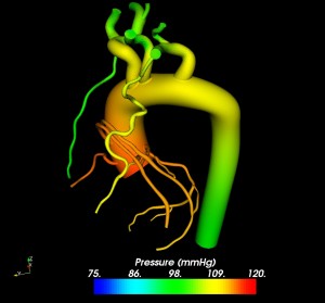
Significant advances are being made in simulations for medical devices. The picture on the left, shared with me by Dr. Tina Morrison, former post-graduate student in Charles Taylor’s group at Stanford University shows the predicted pressures in a patient specific aorta.
 Flow and pressure waves emanate from the health and travel through the major arteries where they are damped, dispersed and reflected due to changes in vessel caliber, tissue properties and branch points. For example you can learn about the coupled multidomain method which was successfully applied to solve the non-linear one-dimensional equations of blood flow with a variety of models of the downstream domain and recently extended to three-dimensional finite element modeling of blood flow and pressure in the major arteries in Comput. Methods Appl. Mech. Engrg. 195 (2006) 3776–3796.
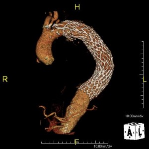
The ability to utilize patient specific medical imaging data depends is revolutionizing the treatment of individual patients and providing a means to better understand the boundary conditions experienced by implantable medical devices. In another image shared with me by Dr. Morrison below a stent is imaged in a descending aorta aneurism. By combining advanced imaging techniques, computational fluid mechanics simulations and Finite Element Analysis (FEA) engineers are taking one step closer to fully describing the mechanics of a medical implant in the human body.


